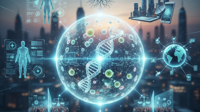
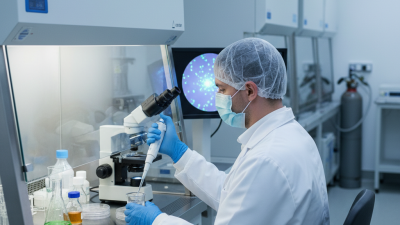
What Is Cell Biology and How Does It Work?
Cell biology explores how living cells are built, supplied, controlled, and repaired. It connects molecular events with visible outcomes, such as muscle contraction, wound healing, or tumor growth. A cell is not a simple container. It is a crowded, responsive system where membranes, proteins, genetic material, and energy pathways interact continuously.
The National Institutes of Health describes cells as the basic units of life, while the National Human Genome Research Institute highlights their central role in interpreting genetic information. These findings support a practical view of biology cell biology: researchers study cells through microscopy, sequencing, cell culture, and biochemical measurement. Under a microscope, a stained cell may reveal a glowing nucleus, moving mitochondria, or damaged membranes. Each image is evidence, but not the whole answer. Experimental design, controls, and repeated measurements remain essential.
The 2024 Nature Index tables show that life-science research remains concentrated among major universities and biomedical institutions worldwide. Industry reports from Grand View Research also identify cell-based research and cell culture as expanding markets, driven by drug discovery, diagnostics, and regenerative medicine. Market forecasts can be useful, yet they are not identical to scientific proof. Their methods and assumptions deserve careful review.
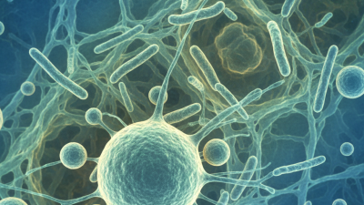
Cell biology works through linked processes rather than isolated parts. Signals cross membranes. Ribosomes build proteins. Mitochondria help manage cellular energy. Errors can spread. A simplified diagram may hide important variation between cells, tissues, and patients. Understanding that uncertainty makes the field more reliable, not less impressive. It also explains why modern medicine increasingly depends on precise cellular evidence.
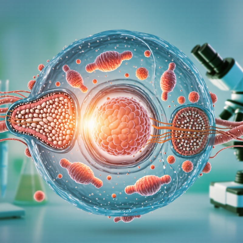
Cell Biology’s Scope: Studying ~37 Trillion Human Cells and Life’s Principles
What Is Cell Biology and How Does It Work? Cell biology examines life at its smallest organized scale: the cell. It asks how membranes control traffic, how genes guide protein production, and how cells cooperate in tissues. A microscope slide may show quiet cells, yet each one exchanges signals, uses energy, and repairs damage.
Cell biology’s scope includes roughly 37 trillion human cells in an average adult body. This estimate comes from a detailed cell-counting analysis by Sender, Fuchs, and Milo in 2016. The study estimated about 3.0 trillion bacterial cells as well. These numbers are estimates, not permanent facts. Body size, age, and counting methods can change them.
The field connects molecules with organs and whole-body health. A muscle cell contracts through coordinated protein movement. A nerve cell sends electrical signals across thin cellular extensions. Immune cells identify danger through receptors on their surfaces. Recent single-cell mapping reports show that tissues contain far more cellular diversity than older diagrams suggested. A neat diagram can mislead. Cells change state, share overlapping roles, and sometimes resist simple labels. Laboratory work must therefore combine microscopy, molecular testing, and statistical analysis. Even the word “cell” hides uncertainty, because some biological boundaries are incomplete or temporary.
What Is Cell Biology and How Does It Work?
Representative diameters of human cell types
Cell biology examines how cells are structured, how they obtain energy, communicate, divide, and specialize. The adult human body contains roughly 30–37 trillion human cells, depending on body size and estimation method. These representative measurements show the wide range of cellular scales, from tiny platelets to the comparatively large human oocyte.
Values are approximate representative diameters. The sperm value refers to the head diameter, while the oocyte value represents its diameter.
Cell Architecture: 7–10 nm Membranes, Organelles, and 3.2-Billion-Base Genomes
Cell biology studies how living cells are built and how their parts cooperate. A cell membrane is often about 7–10 nanometers thick, yet it controls a busy border. Lipids form the flexible barrier, while proteins recognize signals, move nutrients, and remove waste. This thin surface is not merely packaging. It is an active communication system.
Inside, organelles divide difficult tasks into specialized spaces. The nucleus stores genetic information and regulates gene activity. Ribosomes assemble proteins, while mitochondria help produce usable cellular energy. The endoplasmic reticulum folds and transports molecules; the Golgi apparatus modifies and sorts them. Under a microscope, these structures can look like tidy compartments. Real cells are less orderly. Their boundaries shift, collide, and sometimes fail.
A human cell’s haploid genome contains roughly 3.2 billion DNA base pairs. This enormous instruction set fits inside a microscopic nucleus through careful folding around protein structures. Small DNA changes can alter a protein, a pathway, or sometimes nothing noticeable. That uncertainty matters. Biology rarely behaves like a simple machine with one predictable result. Researchers compare imaging, genetic data, and biochemical measurements because any single method can mislead. Even the 7–10 nanometer membrane estimate is a useful range, not a universal rule for every membrane. Understanding cells therefore requires precise measurement, practical caution, and a willingness to revise attractive explanations.
Cellular Chemistry: Enzymes, ATP, and Reactions That Sustain Life
What Is Cell Biology and How Does It Work?
Cellular Chemistry: Enzymes, ATP, and Reactions That Sustain Life
Cell biology examines how tiny living units maintain order, exchange materials, and respond to change. At the center of this work is chemistry. Water surrounds molecules inside cells, while ions and proteins move through crowded spaces. These conditions allow thousands of reactions to happen every second. Without careful control, useful reactions could become harmful or simply stop.
Enzymes act as precise helpers. They bring reactants together and lower the energy needed for a reaction. An enzyme does not disappear after the reaction, so one molecule can assist repeatedly. In a teaching laboratory, a small temperature change can alter enzyme activity noticeably. Too much heat may distort the enzyme’s shape. The reaction then slows. It is a simple demonstration, but real cells face far more variables.
ATP stores chemical energy in a form cells can use quickly. When ATP loses a phosphate group, energy becomes available for transport, movement, and molecule building. Cells often connect this energy release to another reaction that needs energy. This process is called coupling. The model is useful, but incomplete. ATP is not a tiny battery holding energy unchanged; its behavior depends on surrounding molecules, water, and concentration. A cell’s chemistry is therefore less like a machine with fixed parts and more like a restless, regulated network. Small changes matter.
Genetic Information Flow: How 46 Chromosomes Control Protein Synthesis
Cell biology asks how living units preserve information and respond to change. In most human somatic cells, 46 chromosomes sit inside the nucleus. They form 23 pairs, with one chromosome in each pair inherited from each parent. Chromosomes are long DNA molecules wrapped around proteins, not loose instruction cards. That distinction matters. Each gene occupies a defined region, but nearby regulatory DNA helps decide when the gene is active. Skin cells and nerve cells carry nearly the same chromosomes. Their behavior differs because they read different genes.
Protein synthesis begins when a cell copies a selected DNA sequence into messenger RNA. This step is transcription. The RNA leaves the nucleus through tiny nuclear pores and reaches a ribosome. During translation, the ribosome reads three RNA bases at a time. Transfer RNA brings matching amino acids, building a protein chain. The order is precise: DNA to RNA to protein. Yet it is not a simple conveyor belt. Chemical signals, chromatin structure, and RNA processing can alter the message before translation. A single gene may produce several related proteins.
In a teaching laboratory, stained chromosomes appear as compact dark bodies under a microscope. The image is useful, but incomplete. Most genetic activity is too small to see directly. A missing or altered chromosome region can change protein production and development. However, not every DNA change causes illness; its effect depends on location, timing, and cell context. Researchers therefore compare chromosome structure with RNA and protein measurements. I still find the system humbling: 46 chromosomes do not act like 46 independent switches. They communicate through networks, and our explanations remain models that require careful testing.